
Micro-Ultrasound
High-resolution (micro) ultrasound is a prostate imaging technique that operates at a higher transducer frequency than conventional ultrasound. It is used to guide prostate biopsies in real time, and is an area of active clinical research as the field of prostate cancer diagnosis continues to develop.
What is micro-ultrasound?
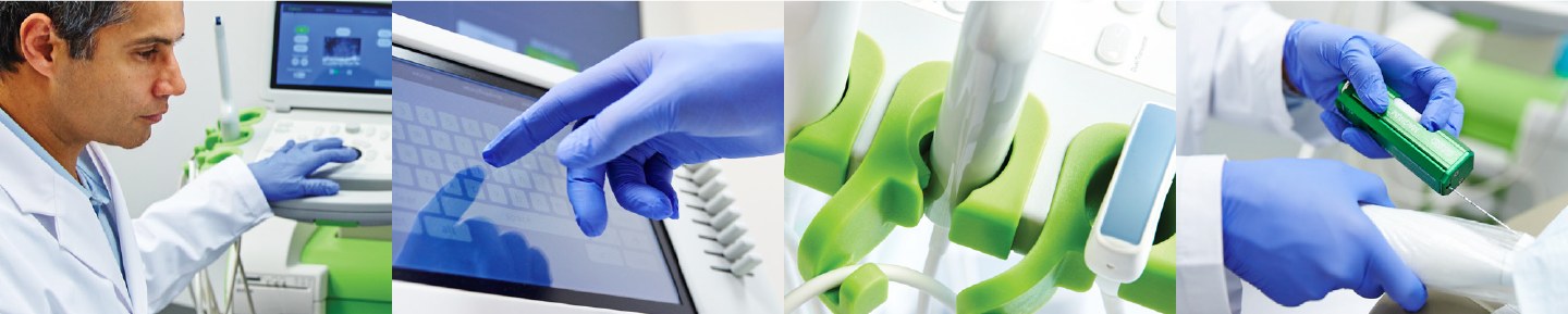
Standard transrectal ultrasound (TRUS) used for prostate biopsy guidance typically operates at 6 to 9 MHz. Micro-ultrasound systems operate at around 29 MHz, which produces images with approximately three times the spatial resolution of conventional ultrasound.1
This higher resolution allows the operator to visualise finer structural details within the prostate - including differences in tissue architecture that are not clearly visible on conventional ultrasound. The images are generated in real time, meaning the operator can see the prostate on screen during the biopsy procedure.
The PRI-MUS risk protocol
A standardised interpretation framework called the Protocol for micro-Ultrasound Risk Stratification (PRI-MUS) was developed to help operators identify and grade suspicious regions on micro-ultrasound images.1 PRI-MUS uses a five-point scale similar in principle to the PI-RADS scoring system used in multiparametric MRI, with higher scores indicating features more likely to be associated with clinically significant prostate cancer.
Published validation work has shown moderate-to-good agreement between operators who have been trained in the protocol, though performance depends on the experience and training of the individual operator.1,2
How does micro-ultrasound compare with multiparametric MRI?
Multiparametric MRI (mpMRI) is currently the best-validated pre-biopsy prostate imaging modality and is recommended in major clinical guidelines, including those of the European Association of Urology (EAU).3 mpMRI is performed before the biopsy, and the images are then used to plan or guide targeted sampling.
Micro-ultrasound operates in real time and does not require a separate pre-biopsy imaging session. Several prospective studies have compared micro-ultrasound-guided targeted biopsy with MRI-guided targeted biopsy, and results have generally shown similar detection rates for clinically significant prostate cancer in selected patient groups.2,4 In 2025, a randomised study (the OPTIMUM trial, published in JAMA) found micro-ultrasound-guided biopsy to be non-inferior to MRI-guided biopsy for the detection of clinically significant prostate cancer.6 However, the total volume of evidence is smaller than for mpMRI, and micro-ultrasound is not yet incorporated into standard guideline pathways as a routine alternative.
Some centres are also investigating the combination of micro-ultrasound guidance with pre-biopsy MRI, using the two modalities together rather than as alternatives.5
Role in targeted prostate biopsy
Prostate biopsy can be performed in two main ways: systematic (a standard grid pattern covering the whole gland) and targeted (directing needles specifically toward areas that appear suspicious on imaging). Most modern biopsy approaches combine both strategies in the same session.
Micro-ultrasound can be used to guide targeted biopsies toward suspicious regions identified during the procedure. Because the guidance is real time, the operator can adjust needle placement in response to what they see on screen. The technique is compatible with both transperineal and transrectal biopsy approaches.
As with all prostate biopsy methods, micro-ultrasound guidance does not eliminate the possibility of missing a cancer, and some regions that appear suspicious on imaging will not contain cancer when the tissue is analysed. Biopsy carries the same general risks regardless of the imaging technology used to guide it, including temporary blood in urine or semen, discomfort, and a small risk of infection.
Micro-ultrasound at Urology NSW - first cases in Australia
In July 2026, Dr Raji Kooner and his team became the first in Australia to use the ExactVu prostate micro-ultrasound. Dr Kooner performed seven consecutive cases, and the team was very happy with the outcomes. We look forward to using this machine on an ongoing basis - it is potentially a significant advance in the management of patients with suspected prostate cancer.
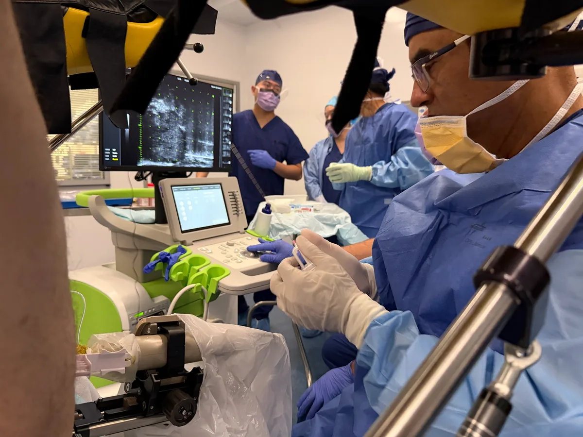
Alongside biopsy guidance, micro-ultrasound may have a role in the active surveillance setting, where patients with prostate cancer are being monitored - this could decrease the need for repeat MRI scans. It is also currently being studied in a primary screening setting, where patients with a borderline PSA test could have this imaging performed. This story is also covered on our news page.
Where micro-ultrasound fits in the diagnostic pathway
For most men with suspected prostate cancer, the standard diagnostic pathway at Urology NSW involves PSA testing, clinical assessment, multiparametric MRI, and - where biopsy is indicated - transperineal biopsy with MRI fusion guidance. Micro-ultrasound represents one of several evolving technologies in this space.
The appropriate imaging and biopsy strategy for any individual depends on clinical history, prior investigations, anatomy, and a detailed discussion of the available options and their relative advantages and limitations. If you have questions about prostate cancer diagnosis, a consultation with a urologist is the appropriate starting point.
Limitations and current evidence status
- Micro-ultrasound is an emerging technology. The published evidence base is growing but remains smaller than for mpMRI.
- Performance is operator-dependent and requires structured training in the PRI-MUS protocol.
- Current major guidelines (EAU, AUA) recommend mpMRI as the standard pre-biopsy imaging pathway; micro-ultrasound is not yet listed as an equivalent alternative in those guidelines.
- The first large randomised controlled trial (OPTIMUM) reported in 2025 and found micro-ultrasound-guided biopsy non-inferior to MRI-guided biopsy for the detection of clinically significant prostate cancer; further studies are ongoing.6
Micro-ultrasound image examples
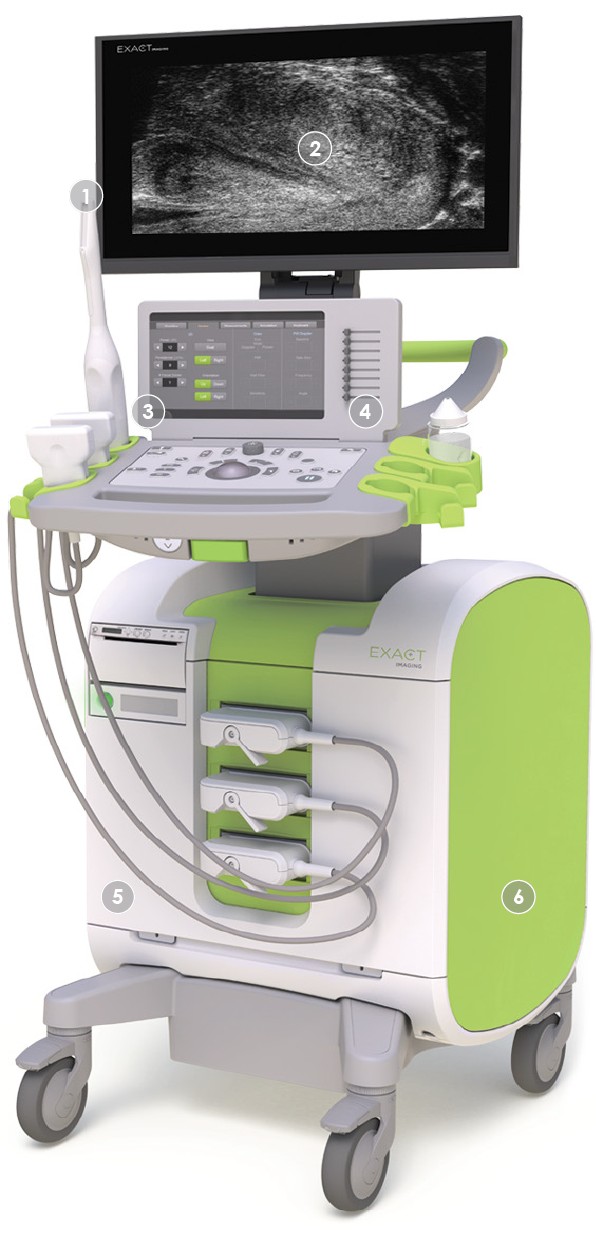
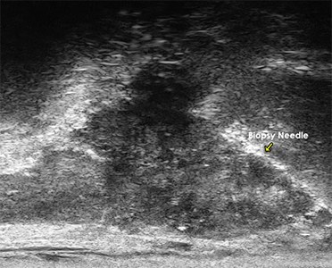
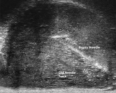
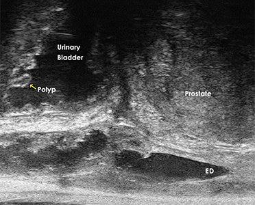
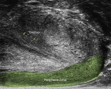
Apart from the photograph of Dr Kooner and his theatre team, images and video on this page are courtesy of Exact Imaging (ExactVu is a trademark of Exact Imaging) and are reproduced with permission. They illustrate the technology only and do not depict any patient of Urology NSW. The availability of a particular imaging technology does not mean it is the appropriate investigation for every person - see the discussion above and talk to your urologist about your individual situation.
References
- Ghai S, Eure G, Fradet V, et al. Assessing Cancer Risk on Novel 29 MHz Micro-Ultrasound Images of the Prostate: Creation and Early Validation of the Micro-Ultrasound Protocol for Prostate Risk Identification. J Urol. 2016;196(2):562-569. doi:10.1016/j.juro.2016.04.073
- Klotz L, Lughezzani G, Maffei D, et al. Comparison of micro-ultrasound and multiparametric magnetic resonance imaging for prostate cancer detection in biopsy-naive men: A multicenter, prospective analysis. Urol Oncol. 2021;39(12):784.e1-784.e9. doi:10.1016/j.urolonc.2021.04.017
- Mottet N, van den Bergh RCN, Briers E, et al. EAU-EANM-ESTRO-ESUR-SIOG Guidelines on Prostate Cancer. European Association of Urology, 2024. Available at: https://uroweb.org/guidelines/prostate-cancer
- Lughezzani G, Saita A, Lazzeri M, et al. Comparison of the Diagnostic Accuracy of Micro-Ultrasound and Magnetic Resonance Imaging/Ultrasound Fusion Targeted Biopsies for the Diagnosis of Clinically Significant Prostate Cancer. Eur Urol Oncol. 2019;2(3):329-332. doi:10.1016/j.euo.2018.09.005
- Abouassaly R, Klein EA, El-Shafei A, et al. Micro-ultrasound versus conventional ultrasound and magnetic resonance imaging for prostate cancer detection: A multi-center, prospective study. Urol Oncol. 2022;40(9):411.e1-411.e8. doi:10.1016/j.urolonc.2022.04.019
- Kinnaird A, Luger F, Cash H, et al. Microultrasonography-Guided vs MRI-Guided Biopsy for Prostate Cancer Diagnosis: The OPTIMUM Randomized Clinical Trial. JAMA. 2025;333(19):1679-1687. doi:10.1001/jama.2025.3579