Transperineal Prostate Biopsy - MRI Fusion
Show video transcript
To diagnose prostate cancer we usually have to do a biopsy of the prostate.
Traditionally we’ve done a transrectal biopsy, which has been through the back passage. The problem with that is it’s associated with an increased chance of infection because you’re clearly going through the bowel.
So over the last 10 or 12 years we developed a different technique called transperineal biopsy, which I’ve been doing.
What we do is, under a light anaesthetic, we put a probe in the back passage and use a little template between the scrotum and the back passage. We put fine needles through that skin between the scrotum and the back passage and take very fine samples of the prostate.
With that, infections are virtually unheard of, and it allows a very accurate pattern of the cancer in the prostate.
These days, with new treatments such as focal therapy where we may look at treating part of the prostate, that very accurate mapping allows us to tell exactly where the cancer is so we can treat it appropriately.
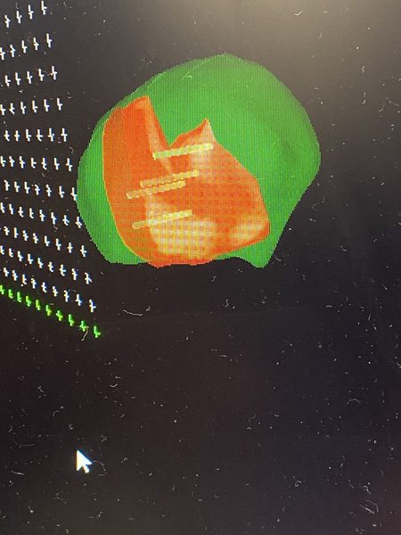
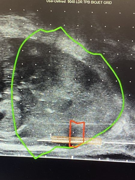
I also use a more sophisticated technique called MRI fusion biopsy. We bring the disc with the MRI into the operating theatre, scan it in, look at the abnormality, and the biopsy is done under ultrasound control.
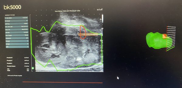
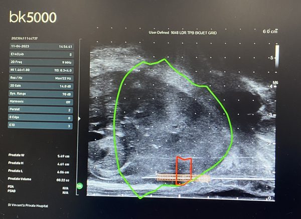
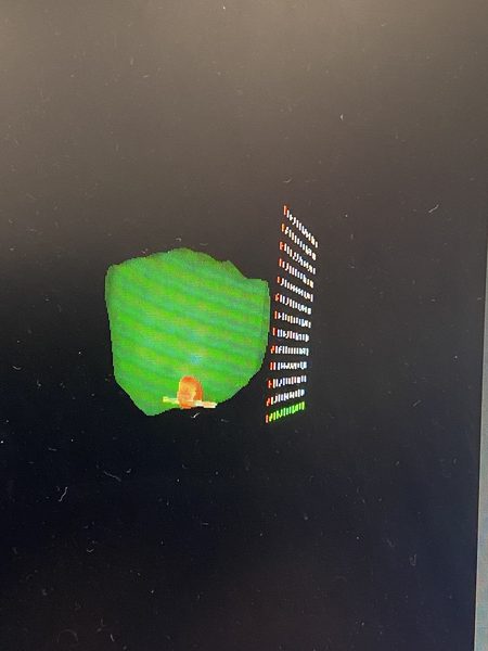
We take the ultrasound pictures and the MRI pictures and overlap them together. In real life we then have a mark on the ultrasound, and we put these fine needles in to take samples exactly in the spot that’s abnormal. That’s called an MRI fusion biopsy, again done by a transperineal method.
These are day cases, patients tolerate them very well, and the risk of infection, as I said, is minimal.
You may have a little bit of blood in the urine, a little bit of blood in the semen, but that’s all self-limiting. Perhaps a slight bit of bruising in the perineum, and that’s self-limiting as well.
So it’s very well tolerated, done under a light anaesthetic, and I think this is a big advance in prostate biopsies.
At Urology NSW, Dr Raji Kooner and his team offer advanced diagnostic techniques for prostate cancer, including the precise and minimally invasive transperineal prostate biopsy. This modern approach provides important advantages over traditional transrectal methods, improving safety and diagnostic accuracy for patients.
What is a transperineal prostate biopsy?
A transperineal prostate biopsy is a diagnostic procedure that involves taking small samples of prostate tissue through the skin between the scrotum and anus (the perineum). These samples are then analysed in a pathology lab to detect the presence of cancerous cells.

Unlike the traditional transrectal ultrasound-guided (TRUS) biopsy, which passes needles through the rectum, the transperineal method avoids contact with the bowel, reducing the risk of infection and sepsis.
Why transperineal over transrectal?
More Australian urologists are now recommending transperineal biopsy due to its clear benefits:
- Lower infection risk. The rectum contains bacteria that can lead to infection. By avoiding the bowel, transperineal biopsy substantially reduces the risk of sepsis, a serious complication that has been reported in up to about 3% of patients after a traditional TRUS biopsy.
- Greater accuracy. This approach offers improved access to all zones of the prostate, particularly the anterior region, which can be missed during a TRUS biopsy. This results in a more accurate diagnosis, especially in men with a rising PSA but previous negative biopsies.
- Better for MRI fusion targeting. Transperineal biopsy is well suited to MRI fusion-guided biopsy, which uses pre-acquired MRI scans to pinpoint suspicious areas with high precision.
- Suitable for local or general anaesthesia. Depending on patient preference and clinical factors, the procedure can be done comfortably under sedation or general anaesthetic.
Who should consider a transperineal biopsy?
At Urology NSW, we may recommend a transperineal biopsy for:
- Men with elevated PSA levels or an abnormal digital rectal examination (DRE).
- Men with previous inconclusive or negative biopsies.
- Men needing a more accurate diagnosis before prostate surgery or radiation.
- Patients seeking a lower-risk, more comprehensive diagnostic pathway.
What to expect during the procedure
- Anaesthesia. The biopsy is usually performed under light sedation or a general anaesthetic for comfort.
- Image guidance. An ultrasound probe is placed in the rectum to visualise the prostate, while needles pass through the perineal skin.
- Tissue sampling. Several samples are taken using a grid or freehand technique. Targeted MRI fusion may be used if pre-biopsy imaging suggests specific areas of concern.
- Recovery. Most patients go home the same day. Temporary minor discomfort or blood in urine or semen may occur but usually resolves within a short time.
MRI fusion biopsy
Dr Kooner has been using MRI fusion technology for prostate biopsies for more than 10 years. The MRI scan performed before the procedure is brought into theatre, and the abnormal area is marked on the scan using a dedicated software program.
The transperineal biopsy is then carried out under ultrasound control. The live ultrasound images are fused with the MRI so that the marked abnormality is overlaid onto the real-time view of the prostate. This allows us to target the area of concern seen on the MRI far more accurately, resulting in a more accurate and precise biopsy.
Dr Kooner also takes great care to use both a cross-sectional and a longitudinal view for every biopsy. Confirming the needle position on two perpendicular planes allows more accurate needle placement and minimises the risk of bleeding or injury to adjacent tissue.
Transperineal biopsy images